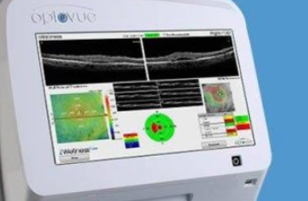
An optical coherence tomography scan (OCT scan) is a critical device for the early diagnosis of many serious eye conditions.
An OCT eye exam is a non-invasive test that provides 3-D color-coded, cross sectional images of the retina to enable early detection and treatment of ocular disease that may develop without any noticeable symptoms.
The OCT scan uses a laser (without radiation) to obtain higher resolution images of the layers of the retina and optic nerve.
The color-coded images provide a wealth of information to help your eye doctor measure the thickness of your retina and identify any optic nerve abnormalities.
The images produced by the OCT scan are also a practical tool in helping patients understand the problem they may be experiencing, as the complication can be seen clearly and in 3D on the screen.
When is an OCT scan used?
OCT scans are used for diagnosis, management or treatment of a variety of ocular conditions:
- Macular edema
- Age-related macular degeneration (AMD)
- Diabetic retinopathy
- Macular hole
- Vitreomacular traction
- Glaucoma
- Drusen
- Retinal occlusions or bleeding
- Retinal detachment
- Retinal pigment epithelium detachment
- Epiretinal membrane
- Retinoschisis
- Central serous chorioretinopathy
- Optic disc edema
- Optic nerve head drusen
- Ganglion cell complex
- Neovascularization
OCT scan for anterior angle examination
The OCT scan is a useful tool in evaluating the anterior angle in patients at risk of developing glaucoma.
The anterior angle is the drainage channel for fluid inside the eye. If the drainage channel is not working properly, it can lead to an increase in eye pressure, and cause damage to the optic nerve and vision loss.
The OCT provides cross-sectional images of the anterior angle, and facilitates detection of open-angle or angle closure glaucoma, as well as narrowing of the anterior angle, which can lead to angle-closure glaucoma.
The OCT scan can also be performed in a dark room, which facilitates faster and more accurate results— especially when the position of the anterior angle in a darkened room is of utmost importance in the diagnosis of angle-closure glaucoma.
Schedule an appointment with an eye doctor near you, if you suspect you may have glaucoma or any other eye condition that an OCT exam can help detect.
SEE RELATED: What Is a Digital Retinal Image?
What should I expect during an OCT exam?
The OCT scan uses light waves to create an image, similar to an ultrasound that uses sound waves to create an image. During the test, you will sit with your chin resting on a support. The OCT will scan each of your eyes, while you fix your gaze on a green target. As the laser scans the retina in each eye, you may notice a red line moving across your vision.
When should I have an OCT exam?
If you are over age 25, or at risk of developing ocular disease, your eye doctor may recommend an OCT exam as part of your annual eye exam. Having regular OCT scans will enable your doctor to compare the images from previous years to detect any abnormalities or thickening of retinal layers.
If you have been diagnosed with an ocular disease, an OCT scan will also provide the information needed to monitor treatment.
If you have any questions or concerns about your OCT exam, never hesitate to speak with an eye doctor near you.
LEARN MORE: Guide to Eye Exams
An OCT exam, as it can facilitate early diagnosis and treatment of sight-threatening ocular diseases— preventing vision loss before symptoms begin.