In the early stages, macular degeneration may not present with noticeable symptoms— a comprehensive eye exam is the only way to detect this serious condition.
Eye exams aren’t just to check your eyesight, they’re an integral part of maintaining eye health, so you can enjoy a life of clear vision.
Annual eye exams allow your doctor to detect changes in the retina and macula— facilitating early diagnosis and treatment of macular degeneration (AMD), along with other sight-threatening eye diseases.
The following tests may be conducted during an eye exam to identify any structural or visual changes:
Amsler grid
An Amsler grid contains straight black lines with a large dot in the center.
The eye doctor will ask you to look at the black dot and point out any irregularities on the grid, such as black spots, or lines that appear:
- Blurry
- Wavy
- Broken
- Faded
- Fractured
Dilated eye exam
Dilating your eyes with drops allows your doctor to more easily and efficiently inspect the back of your eye, where the retina and macula, the center of the retina, are located.
Using a slit lamp with high magnification, your doctor will look for drusen, yellowish deposits that grow beneath the retina. These deposits create a speckled appearance and are often the first signs of AMD.
Other retinal irregularities may include pigment changes, abnormal blood vessels or fluid leakage within the macula.
For a comprehensive macula examination schedule a visit with an eye doctor near you.
SEE RELATED: The Importance of Pupil Exams
Digital retinal imaging (DRI)
Digital retinal imaging has become a vitally important test, used for early detection of retinal diseases.
Digital retinal imaging, also known as a retinal photograph, is a non-invasive diagnostic tool used for early detection of retinal diseases. This imaging technique produces high resolution, colored images of the retina, optic nerve, and blood vessels located in the back of the eye.
With DRI, your doctor can more easily detect and track changes to your ocular health and vision.
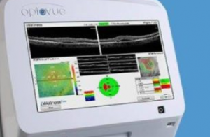
Optical coherence tomography (OCT)
The OCT is a non-invasive imaging tool that produces detailed 3D, cross-sectional images of the retina.
These 3D full color images are very effective in picking up the earliest signs of retinal and macula changes, before most other devices. These images can detect thinning, thickening or swelling of the macula and retina, caused by leaking blood vessels in and under the retina.
Fluorescein angiography (FA)
Fluorescein angiography (FA) is a diagnostic test used for early detection of AMD, diabetic retinopathy and other retinal conditions.
During this test, fluorescent dye is injected into a vein in the arm. As the dye flows through the blood vessels in the eye, any irregularities are highlighted by the dye.
In addition, a unique camera captures images to help further identify any abnormalities within the blood vessels or retina.
Indocyanine green angiography (ICGA)
The indocyanine green angiography (ICGA), similar to FA, involves an injected dye, and is often used in conjunction with FA to pinpoint the type of macular degeneration detected.
The ICGA is used to examine the blood flow in the choroid, the layer under the retina, and provides a better understanding of the health of these sub-retinal blood vessels.
LEARN MORE: Guide to Eye Exams
For a comprehensive macula examination schedule a visit with an eye doctor near you.
Eye exams aren’t just for identifying refractive errors, they can also check for eye diseases, such as macular degeneration (AMD).
The earlier AMD is detected, the greater your chances of preserving your clear vision and enjoying the best quality of life.