Many ocular diseases develop without any noticeable symptoms.
Advanced diagnostic equipment allows doctors to detect signs of ocular disease before symptoms are present —preventing significant and permanent vision loss, and enabling early treatment to manage the disease.
Corneal Mapping (Topography)
Corneal Mapping, also known as Corneal Topography, is a non-invasive diagnostic tool that provides 3-D images of the cornea (the outer layer of the eye responsible for about 70% of the eye’s focusing power). The images provide details about the cornea’s shape and curvature, and enable detection of irregular conditions.
Corneal mapping is used to detect certain corneal diseases, abrasions, deformities, and irregular astigmatisms. Corneal mapping is also used to monitor and treat ocular conditions, plan eye surgery, and fit contact lenses.
The diagnostic test involves sitting in front of a lighted “bowl” and resting your head against a bar. After your data is collected, a color coded image of the corneal shape is generated, using different colors to differentiate elevations (similar to a topographic map of the earth that displays changes in the surface of the land).
Digital Retinal Imaging
Digital Retinal Imaging is a non-invasive, diagnostic tool that produces high resolution images of your retina, optic nerve, and other retinal structures at the back of your eye. These retinal images facilitate early detection of ocular diseases, such as glaucoma, diabetic retinopathy and macular degeneration.
The images are stored electronically to allow your doctor to detect and measure any changes to your retina at each eye exam.
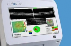
Optical Coherence Tomography (OCT)
An Optical Coherence Tomography scan (commonly referred to as an OCT scan) is a non-invasive, diagnostic tool that provides color-coded, cross sectional images of the retina, allowing for early detection and treatment of ocular disease.
The OCT scan uses a laser (without radiation) to obtain higher resolution images of the layers of the retina and optic nerve.
It is a critical tool for early diagnosis of macular degeneration, glaucoma, retinal detachment, and diabetic retinal disease.
If you suspect you have an eye condition, contact an eye doctor near you, who can diagnose and treat the condition.
SEE RELATED: Eye Exams
Visual Field Testing
A Visual Field test is a diagnostic test that produces a computerized map of your visual field. It enables your doctor to measure the range of your peripheral vision, or detect any abnormalities in your visual field.
During an initial screening, you will be asked to describe what you see, while maintaining a fixed gaze on a central object, and covering one eye. During a more comprehensive assessment, you will look into a special machine and press a button when you see a flash of light. At different stages of the test, the lights are brighter or dimmer. Each eye is tested separately.
After the test, your doctor will examine the map and determine any visual field disturbances.
OPTOS Retinal Exam
The Optomap® Retinal Exam enables a wide view of the retina, producing a highly detailed image for early detection of ocular diseases (such as, macular degeneration, glaucoma, retinal tears or detachments).
Using the optomap®, your eye doctor can also detect signs of diabetes, hypertension, or other health related issues. During the optomap® retinal exam an image of your retina is captured, and immediately displayed on a computer screen. This allows for you to view and discuss with your doctor, any changes that may have been detected.
The image is stored electronically to allow your doctor to detect and measure any changes to your retina at each eye exam.
LEARN MORE: Guide to Eye Exams
Schedule an appointment with an eye doctor for a comprehensive eye exam, and to discuss any questions you may have about your eyes or eye health.