Glaucoma is a progressive ocular disease that causes vision loss from damage to the optic nerve, which is responsible for carrying visual signals between the eye and brain.
- Glaucoma is the #1 cause of irreversible blindness, worldwide
- Glaucoma accounts for 12.3% of global blindness.
- Glaucoma affects up to 5% of the people aged 70+ years and increases to over 9% for those aged 80+ years.
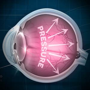
The most common form of glaucoma occurs when there is high pressure inside the eye (intraocular pressure, IOP), caused by a build-up of fluid that cannot drain properly.
Why is glaucoma called the “Silent Thief of Sight”?
This nickname is well deserved as glaucoma causes vision loss before the person notices any symptoms. Glaucoma has the infamous reputation of “robbing” the ability to see, without giving the person time to recognize symptoms and seek their eye doctor’s medical opinion. Consequently, a diagnosis is usually made only after permanent vision loss as occurred.
It is therefore important to schedule regular eye exams, as new diagnostic tests are available to detect early signs of glaucoma, and are essential in preventing vision loss.
What is ‘tunnel vision’?
The damage to the optic nerve affects peripheral vision first. If glaucoma is not effectively controlled at an early stage, the disease can cause severe peripheral vision loss— resulting in a condition called ‘tunnel vision’. Tunnel vision causes the ability to use your ‘side vision’, and limits your field of vision to strictly seeing images that are in your central vision, or straight ahead.
Imagine looking through a paper tube— this is what living life with ‘tunnel vision’ is like.
As glaucoma progresses into a more advanced stage, the disease begins to affect central vision as well— resulting in partial or complete permanent vision loss.
Types of glaucoma
There are a few different types of glaucoma:
- Chronic (open angle) glaucoma is the most common form, caused by IOP build-up over time, typically as a result of aging. About 2.7 million Americans, above the age of 40, are affected by open-angle glaucoma.
- Acute (angle closure) glaucoma is a rare form that requires immediate medical attention. It develops as a result of sudden pressure build-up. Symptoms include blurred vision, seeing halos around lights, eye pain, headaches, nausea and vomiting. About 1 in 1,000 people develop acute glaucoma in their lifetime.
- Secondary glaucoma can develop from complications of medical conditions (i.e. diabetes and hypertension), other ocular conditions, side effects of medications, or trauma to the eye. Secondary glaucoma accounts for 10% of all glaucoma cases.
- Normal tension glaucoma develops when the optic nerve is damaged, without a presence of high IOP. The cause of this type of glaucoma is still unknown. Normal tension glaucoma occurs in approximately 30-40% of all patients diagnosed with glaucoma.
Schedule an eye exam with an eye doctor near you who has experience diagnosing and managing glaucoma.
SEE RELATED: Ocular Hypertension
Risk factors and prevention
Certain factors can increase your risk of developing glaucoma:
- Over age 40
- Family history of glaucoma
- Ancestry of African American, Hispanic, Asian and Native American or Indigenous Canadian descent
- Previous eye injury, trauma, or surgery
- Thinning of cornea
- High myopia (nearsightedness)
- Prolonged use of steroids
- Diabetes
- Hypertension
Diabetes and hypertension are risk factors that you may be able to control. However, as most risk factors are genetic, it is crucial that you schedule regular eye exams, and inform your doctor of any factors that put you at a higher risk of glaucoma.
Diagnosing glaucoma
Diagnosing glaucoma requires the skill and experience of an eye doctor, and specific examination tools.
Tonometry is a specialized test that measures intraocular pressure (IOP). This test is usually performed by a non-contact device which delivers a puff of air toward the eye. This device will calculate your IOP based on your eye’s response to the air. The benefit of this device is that local anesthetic eye drops are not required.
To obtain another measure of your IOP, yellow numbing drops will be placed into your eyes so that your doctor can gently touch the surface of your eye with either a tonometer or tonopen. If you have high IOP, you may have, or be at risk for glaucoma.
An optical coherence tomography (OCT) scan is a non-invasive, diagnostic tool that provides color-coded, cross sectional images of the retina, allowing for early detection and treatment of glaucoma, and other ocular diseases. The OCT scan uses a laser (without radiation) to obtain high resolution images of the layers of the retina and optic nerve to locate any changes or ocular damage.
A visual field test is a diagnostic test that produces a computerized map of your visual field. It enables your doctor to measure the range of your peripheral vision, and detect any abnormalities in your visual field.
During an initial screening, you will be asked to describe what you see, while maintaining a fixed gaze on a central object, and covering one eye.
During a more comprehensive assessment, you will look into a special machine and press a button when you see a flash of light. At different stages of the test, the lights are brighter or dimmer. Each eye is tested separately. After the test, your doctor will examine the map and determine any visual field disturbances.
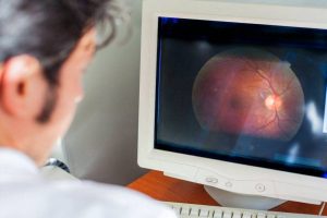
Optic disc examination involves a thorough evaluation of the optic disc, the surface of the optic nerves, which carries the visual information from the eyes to the brain. Your eye doctor will examine the quality of the margin, or neuroretinal rim, and optic disc cup.
This examination can be performed with direct ophthalmoscopy, slit lamp, optic disc photography, or with digital imaging devices.
Examination of anterior angle (Gonioscopy) enables a thorough evaluation of the internal drainage system of the eye, or the anterior chamber angle— where the cornea and the iris meet.
Fluid from inside the eye (aqueous humor) drains out of the eye through this angle, and into the veins at the back of the eye. Through this test, your eye doctor will be able to determine if the angle is open or closed, and detect the presence of abnormal blood vessels, adhesions (synechiae), or damage from previous eye trauma.
To enable a clear view of the angle and its drainage system, your eye doctor will place a special contact lens prism on the surface of your eye after inserting numbing eye drops. This test is relatively quick, and does not cause any pain, though you may feel some pressure from the contact lens.
When to see an eye doctor
If you or a loved one is experiencing symptoms of glaucoma, schedule a comprehensive eye exam for a proper diagnosis.
LEARN MORE: Guide to Eye Conditions
With early detection of glaucoma, clear and comfortable vision can be achieved— with an increased chance of preserving your vision and quality of life.