Retinal artery occlusion (RAO) is an eye emergency and causes sudden vision loss in over 2 people per 100,000 every year.
The retinal nerve layer inside the eye receives oxygen from the retinal artery, which enters the eye through the optic nerve.
What is retinal artery occlusion (RAO)?
RAO is a type of retinal disease occurs when there is blockage of the retinal artery which carries oxygen to the nerve cells in the retina at the back of the eye.
A lack of oxygen flow to the retina will cause a sudden retinal disease which is an emergency due dramatic vision loss.
There are two types of RAOs:
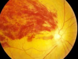
- Central retinal artery occlusion (CRAO) is a blockage in the central artery in your retina.
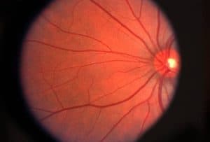
- Branch retinal artery occlusion (BRAO) blocks the small arteries, that branch off the central artery.
What causes RAO?
RAO occurs when an embolus, a tiny particle of cholesterol that restricts blood flow, or thrombus, a blood clot, obstructs the retinal artery – a serious retinal complication.
If the blockage breaks up and blood flow to the retina is restored, the retinal artery occlusion may be transitory, lasting only a few seconds or minutes, however if the blockage remains the vision loss will be permanent.
Common risk factors for RAO include:
- Abnormal heart rhythms such as atrial fibrillation
- Atherosclerosis (fatty deposits in the arteries)
- Blood platelet abnormalities
- Carotid artery disease
- Diabetes
- Disorders contributing to blood clot formation, such as sickle cell disease
- Faulty heart valves (valvular heart disease)
- Giant-cell arteritis
- High blood pressure
- Intravenous drug abuse
- Pregnancy
- Tumors in the heart (myxoma)
- Use of oral contraceptives
- Homocystinuria (a hereditary disorder that prevents your body from processing the amino acid methionine)
What are retinal artery occlusion symptoms?
The obstruction of a retinal artery is frequently accompanied by a painless loss of vision in one eye. The visual loss is determined by the area of the retina impacted by the blocked arteries.
Symptoms are depending on the type of RAO.
- CRAO causes sudden and severe vision loss.
Fortunately, 25% of patients with CRAO have an extra artery in their eyes called the cilioretinal artery. When CRAO strikes, having a cilioretinal artery can significantly reduce the risk of central vision impairment, as long as the cilioretinal artery is unaffected.
- BRAO may result in partial vision loss in your visual field.
BRAO may go unnoticed with no symptoms if the afflicted area is not in the center of the eye or is very limited in size.
SEE RELATED: Guide to Retinal Diseases
If you’ve experienced any sudden vision loss, contact an eye doctor near you.
How is RAO diagnosed?
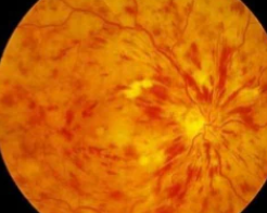
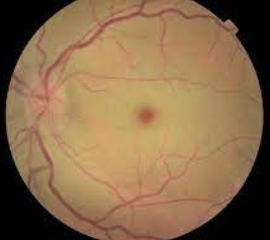
To diagnose CRAO, a dilated eye examination typically reveals a ‘cherry red spot’ appearance of the retina.
The red spot appears as the macula appears bright red, but the surrounding retina appears pale, due to a lack of blood flow.
BRAO appears as an area of superficial retinal whitening along with obstructed vessels.
The whitening of the retina lasts about 4 to 6 weeks until it starts to fade. The filling of the retinal arteries is delayed, according to fluorescein angiography (FA).
Optical coherence tomography (OCT) reveals swelling in the inner layers of the retina in the affected area, which atrophy over time and become much thinner than normal.
RAO Treatment
There is no clinically proven treatment for CRAO; however there are several therapies that may be used including:
- Hyperventilation – inhaling carbogen, a combination of 95% oxygen and 5% carbon dioxide, in an attempt to widen the retinal arteries and remove the clot.
- Paracentesis – the removal of fluid from the front of the eye with a small-gauge needle to try and remove the embolus. This is done to lower the intraocular pressure.
- Medication – to lower the intraocular pressure.
- Ocular massage – to dislodge the clot
- Intravitreal injections – usually anti-VEGF medications.
- Laser photocoagulation therapy – to try to reduce the oxygen demand of the retina and thus stop the abnormal blood vessels from developing. The laser is used to create tiny burns in the retina, usually near the area of the occluded artery.
However, in order for any treatment to be effective in CRAO, it must be used within a limited time frame, most likely within 4 to 6 hours after the onset of symptoms.
Unfortunately, none of these treatments has been shown to modify disease progression in a predictable way.
LEARN MORE: Guide to Eye Health
If you experience any sudden loss of vision, schedule an appointment with an eye doctor near you who can evaluate your eyes and vision.
A person suspected of having sudden vision loss or any retinal condition should be rushed to the nearest emergency room or to a primary care physician as soon as possible.
Retinal artery occlusion is an urgent medical emergency.