Over 2.5 million adults are affected by CRVO, every year.
The central retail vein drains all the blood from the retina, carry away carbon dioxide and waste products. Any damage to this vein can cause sudden retinal disease and dramatic vision loss and blindness.
Central retinal vein occlusion (CRVO)
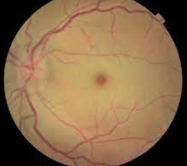
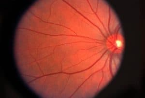
Central retinal vein occlusion (CRVO) is a serious retinal condition in which the main vein that drains blood from the retina becomes blocked or closes off partially or completely.
There are two types of CRVO:
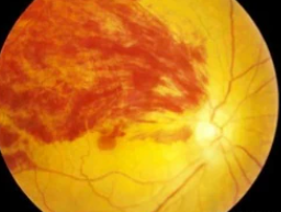
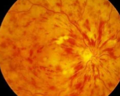
- Non-ischemic CRVO – a mild type characterized by leaky retinal vessels with macular edema
- Ischemic CRVO—a more severe type with closed-off small retinal blood vessels
What causes CRVO?
CRVO affects one eye in the majority of patients. CRVO has no established etiology, despite the fact that diabetes and high blood pressure are risk factors.
What we do know is that CRVO is caused by a blood clot or decreased blood flow in the retina’s central retinal vein.
CRVO that occurs in both eyes at the same time can be related to systemic disease. If this is the case, a tendency toward irregular blood clotting is definitely more common and medical testing to detect thrombophilia (an increased tendency to develop blood clots) is indicated.
Symptoms of CRVO
Non-ischemic CRVO may show no symptoms. However, many patients with CRVO have symptoms such as blurry or distorted vision due to swelling of the macula (center part of the retina). Some patients have mild symptoms that appear and disappear, called transient visual obscurations.
Patients with ischemic CRVO have worse vision and have a lower likelihood of recovery.
These patients have a tendency to generate new blood vessels in the eye, which can obstruct the outflow of normal ocular fluids in the front of the eye.
Glaucoma may also develop as the ocular pressure rises. New blood vessels in the rear of the eye may produce bleeding. Patients may also experience pain, redness, and irritation.
SEE RELATED: Guide to Retinal Disease
If you’ve experienced sudden loss of vision or eye pain, contact an eye doctor near you.
How is CRVO diagnosed?
CRVO is usually diagnosed clinically, based on medical indications and symptoms described by the patient.
When a retina specialist examines the back of the eye, using digital imaging, a distinct pattern of retinal hemorrhages is visible, and a diagnosis is made.
Diabetic retinopathy and retinopathy caused by low blood counts, such as anemia and thrombocytopenia (a deficiency of blood platelets), are two common diseases that can mimic CRVO.
Macular edema, or swelling of the center of the retina, is common, and an optical coherence tomography (OCT) scan is frequently obtained to detect this and assess the extent of swelling.
Fluorescein angiography (FA) imaging may be used to help distinguish CRVO from disorders that are similar to it, as well as to assess the closure of small blood vessels and to look for or confirm the formation of new abnormal vessels.
CRVO treatment
About one-third of older patients who receive no treatment improve on their own, one-third wax and wane and stay about the same, and one-third deteriorate..
The vascular endothelial growth factor (VEGF) is raised in people with CRVO, which causes swelling and the formation of new vessels that are prone to bleeding.
The most popular treatment, which is based on the outcomes of large randomized clinical trials, involves injecting an anti-VEGF medicine into the eye on a regular basis to prevent new blood vessel formation and swelling.
Another option for treating macular edema from CRVO is with an injection of intraocular steroid.
LEARN MORE: Guide to Eye Health
Schedule an appointment with an eye doctor near you who can diagnose and treat CRVO, early treatment might save your vision.
CRVO is a serious eye condition, requiring urgent medical care.
Early diagnosis of macular edema or abnormal blood vessels is critical; most people can prevent serious vision loss if treatment is started before significant damage to the eye occurs.