Over 33% of patients with pigment dispersion syndrome will develop high eye pressure or glaucoma.
Learn about a type of glaucoma that is caused by pigment granules flaking off from the iris, clogging the eye’s drainage system and causing excessive eye pressure and damage to the optic nerve.
What is pigment dispersion syndrome (PDS)?
Pigment dispersion syndrome (PDS) causes pigment to be released in the eye. These pigment deposits can clog the eye’s drainage system, resulting in increased ocular pressure.
What is pigmentary glaucoma?
Pigmentary glaucoma is a type of secondary open-angle glaucoma characterized by trabecular meshwork pigmentation. Pigment in this area can block the natural drainage channel of fluid to outside the eye.
The blockage of the channel will lead to increased eye pressure and the serious sight-threatening eye condition, glaucoma.
When the pigment dispersion syndrome causes damage to the optic nerve, pigmentary glaucoma (PG) is diagnosed.
Not all patients that have pigment dispersion will develop glaucoma.
What causes the release of eye pigment?
It is thought that due to the anatomy of the eye, there’s friction or contact between the iris and the lens, causing chafing or rubbing between these structures during regular activities like reading.
It’s also possible that strenuous activity causes an increase in pigment release. As a result, high-risk individuals are prescribed eye pressure medications prior to activity or to avoid exercise altogether.
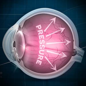
Why does the eye pressure increase?
Fluid inside the eye drains out through a drainage channel, known as the anterior angle.
If the drainage system is unable to cope with the increased load, the ocular pressure rises — eventually leading to glaucoma.
The pigment that is discharged can obstruct the drainage system causing problems with the fluid outflow, leading to increased pressure inside the eye.
Some people’s drainage systems can handle the excess pigment without any issues, and glaucoma doesn’t develop.
SEE RELATED: What is Glaucoma?
Contact an eye doctor near you who can detect and diagnose glaucoma.
Pigmentary glaucoma is under-diagnosed
Pigmentary glaucoma is frequently underdiagnosed due to the lack of symptoms and the fact that it usually develops at a younger age than other forms of glaucoma.
Patients with this type of glaucoma frequently do well if diagnosed early and treated effectively.
There is a chance that the pigment dispersion will fade over time, and that eye pressure will return to normal levels in some cases.
How is pigmentary glaucoma diagnosed?
Many individuals, especially early in the disease process, don’t show early symptoms.
When pigmentary glaucoma progresses, patients may develop side vision problems, similar to those seen in open-angle glaucoma.
Patients may see halos or have blurry vision on a sporadic basis.
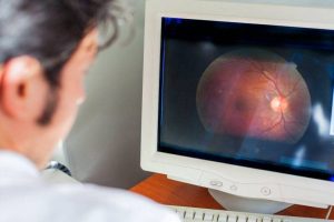
An eye doctor will perform a complete examination of your eyes, including an evaluation of your drainage angle, in order to identify pigmentary glaucoma.
Pigment dispersion or pigmentary glaucoma patients have more pigment in the drainage angle than normal. They may also have pigment on the inner lining of the cornea, as well as thinning of the iris, which causes iris chafing and the release of pigment.
A thorough examination of the rear of the eye is required to detect optic nerve damage. To check for side vision abnormalities, your eye doctor may image your optic nerve and perform field of vision tests.
How to treatment pigmentary glaucoma
The treatment for pigmentary glaucoma is similar to the treatment for open-angle glaucoma in that it entails decreasing eye pressure using eye drops, medicines, a laser procedure or glaucoma surgery.
Medication
Pigmentary glaucoma is treated with the same eye drops or drugs that are used to treat open-angle glaucoma.
Laser or Eye Surgery
A laser procedure to create a small hole in the iris is commonly used to prevent or treat angle-closure glaucoma, and may be used for pigmentary glaucoma.
The objective of the hole in pigmentary glaucoma is to help shift the architecture in the eye, resulting in less chafing or rubbing of the iris and hence less pigment dispersion.
Because of the increased pigment in the drainage angle in pigmentary glaucoma, a new type of laser (laser trabeculoplasty) can also be utilized to improve drainage and lower pressure.
For more aggressive forms of pigmentary glaucoma, trabeculectomy or tube shunt surgery may offer the best outcome.
Regular follow-up checkups with your eye doctor are crucial to the preservation of sight in any kind of glaucoma.
LEARN MORE: Guide to Eye Conditions
Schedule an appointment with an eye doctor near you to have your eye pressure checked.
Pigmentary glaucoma is caused by pigment granules flaking off from the iris, clogging the eye’s drainage system and causing excessive eye pressure and damage to the optic nerve.
Early diagnosis is vital to achieve the best results.