Pingueculae and pterygia are benign growths that appear on the eye’s conjunctiva, the clear covering over the white part of your eye.
What is a pinguecula?
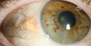
A pinguecula (plural pingueculae) is a small, yellowish raised growth, that develops with a triangular protrusion near the cornea. It usually develops on the side of the eye near your nose, but can grow on the other side too. A pinguecula is usually small in size but can grow larger over time— though this is rare.
It can develop at any age, but tends to be more common in middle aged adults.
What causes a pinguecula to develop?
A pinguecula occurs when deposits containing protein, fat and/or calcium, form on the tissue of the conjunctiva.
The exact cause of pinguecula has yet to be discovered, but there is a correlation between excessive unprotected exposure to sunlight, wind, dryness and dust.
Symptoms of pinguecula
A pinguecula may not cause any symptoms, or may cause the following mild symptoms:
- Dryness
- Irritation
- Foreign body sensation
In more severe cases, you may experience:
- Inflammation
- Itching
- Redness
- Soreness
How is a pinguecula treated?
In most cases, no treatment is necessary— other than to protect your eye from overexposure to the sun and other harmful elements.
However, if the pinguecula is causing discomfort, there are treatments available to alleviate your symptoms.
Eye drops or ointments can help to relieve dryness, irritation and itching.
Steroid eye drops with anti-inflammatory medication may be prescribed for swelling.
In rare cases, surgery to remove the growth may be recommended if the pinguecula is causing serious vision problems or preventing blinking.
If you’re experiencing any eye pain or discomfort, contact an eye doctor near you.
SEE RELATED: What Is a Pinguecula?
What is a pterygium?
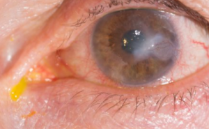
A pterygium is a growth of fleshy tissue containing blood vessels which may cause it to appear pink sometimes, though it typically has a white flesh-colored appearance, with a round, oval, or anvil shape.
A pterygium may develop in one or both eyes, and typically grows in the corner of the eye— near the nose and toward the cornea. A pterygium develops on the surface of the cornea, and is more likely to grow over the cornea than a pinguecula.
A pterygium often starts out as a pinguecula, until it begins to grow over the cornea. It is also more visible than a pinguecula.
A pterygium may remain small in size, or grow large enough to cover part of the cornea— leading to vision problems.
What causes a pterygium to develop?
Similar to a pinguecula, a pterygium (plural pterygia) is believed to be caused by prolonged unprotected exposure to UV rays from the sun, and are sometimes called “surfer’s eye”.
They are most common in middle-aged adults between ages 20 to 50, who live in sunny, dry climates and spend a significant amount of time outdoors.
Symptoms of pterygium
In most cases, there are no symptoms.
However, some people may experience mild symptoms similar to those of a pinguecula:
- Dry eyes
- Redness
- Irritation
- Inflammation
- Foreign body sensation
- Contact lens discomfort
In severe cases, the pterygium may grow far enough into the cornea and obstruct vision, or cause the cornea to change shape— resulting in astigmatism.
How is a pterygium treated?
If necessary, treatment for symptoms of a pterygium may be similar to those used for pinguecula— lubricating or steroidal eye drops or ointments.
Surgery is more common for a pterygium because of its potential for vision changes.
Surgery will be recommended when a pterygium is causing vision loss or an astigmatism, which can cause blurry vision. Your eye doctor may also recommend surgery to remove the pterygium if eye drops or ointments are not providing relief of your symptoms.
Some patients may opt for surgery for cosmetic reasons.
In some cases, a conjunctival graft is performed to prevent recurrence— which can happen. During this procedure, a small piece of tissue is grafted onto the area where the pterygium was removed.
How are these conditions diagnosed?
Your eye doctor can diagnose these conditions through a comprehensive examination, using a slit lamp. This lamp enables a clear view of your eye with the help of bright lighting and magnification.
Additional tests may include:
- Visual acuity test involves reading letters on an eye chart to assess clarity of distance vision.
- Corneal topography is a medical mapping tool used to measure any curvature changes in your cornea.
- Photo documentation involves taking pictures to track and record the growth rate of the pterygium.
Prevention
- Avoid prolonged exposure to sun, wind, dust
- Wear sunglasses with UV protection
- Wear protective eyewear in dry and dusty environments
- Wear a hat for extra protection
- Keep your eyes moisturized with artificial tears
LEARN MORE: Guide to Corneal Conditions
If you think you may have a pterygium or pinguecula, schedule an exam with an eye doctor for a proper diagnosis and appropriate treatment plan.
While a pterygium and pinguecula are often completely benign conditions, they should be closely monitored by an eye doctor to ensure that they do not progress into a more serious condition which may threaten your vision.