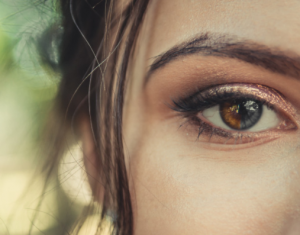
Approximately 80 percent of everything we learn comes through our eyes — the question is, how?
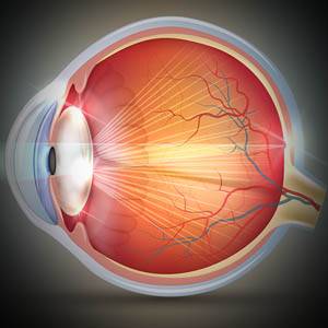
The eye contains over two million working parts and is considered the second most complex organ in the body— the most complex is the brain.
The inner structures of the eye all work together to produce an image that your brain can understand.
In order to produce a clear image, the eyes must complete a five step process:
Step 1: Light enters the eye through the cornea
When we look at an object, the light that is reflected off of the object enters the eye through the clear front layer of the eye, called the cornea. The cornea bends the light before it passes through a watery substance that fills the area behind the cornea, called the aqueous humor.
Step 2: The pupil adjusts in response to the light
The light continues to travel through the black opening in the center of the iris, called the pupil. The iris is the colorful part of your eye that gives it its blue, green, hazel, brown or dark appearance.
The pupil then automatically gets bigger or smaller, depending on the intensity of the light.
How does the pupil expand and contract?
The iris is actually made up of muscles that expand and contract to control the pupil and adjust its size. So when you see your pupil getting bigger or smaller, it is really the iris that is controlling the pupil opening in response to the intensity of light entering the eye.
Step 3: The lens focuses the light onto the retina
The light passes through the pupil to the lens behind it. The lens adjusts its shape to bend and focus the light a second time, to ensure that you have a clear image of what you are looking at.
At this point, the light has been bent twice— as it moved from the cornea through the lens, and then from the lens to the retina. This “double bending” has actually flipped the image upside down.
If you suspect you have blurry vision or an eye condition, contact an eye doctor near you, who can diagnose and treat the condition.
SEE RELATED: Eye Anatomy: The Front of the Eye
Step 4: The light is focused onto the retina
The light then passes from the lens to the back of the eye which is filled with a clear, gelatinous substance called the vitreous until it reaches the retina, the light-sensitive layer at the back of the eye.
The light is then focused throughout the retina which contains nerves called photoreceptors. The photoreceptors are made up of rods and cones, and are responsible for transforming the light rays into electrical impulses. While the light is focused throughout the retina, most of the light entering the eye is focused onto the focal point on the retina, known as the macula.
Step 5: The optic nerve transmits visual information to the brain
The nerves of the retina collect all of the electrical impulses, which then travel through the optic nerve at the very back of the eye up to the occipital lobe in the back of the brain.
At this point, The light then passes from the lens to the back of the eye which is filled with a clear, gelatinous substance called the vitreous until it reaches the retina, the light-sensitive layer at the back of the eye.
The eye-brain connection
Vision is dependent on the connections between the eyes and the brain.
The light that enters the eye is required to go through a specific process in order to focus properly on the retina.
If the connections between the eye and brain are not well developed, the visual information that is sent to the brain will not be interpreted properly, and the image will be difficult to see.
The eye in its perfection
The process of seeing is dependent on the perfection of the eye and all of its components, including:
- Eyeball shape
- Corneal shape and integrity
- Lens clarity and curvature
- Retinal health
If any of these components do not function properly, or are irregularly shaped, vision problems can occur— most commonly, blurry vision will develop.
When this happens, corrective lenses in the form of eyeglasses or contact lenses are prescribed to help the light focus accurately onto the retina and enable clear vision.
Parts of the eye
Cornea: The clear dome-like structure that covers the front of the eye and is responsible for bending light as it enters the eye.
Pupil: The dark opening in the center of the eye that opens and closes in response to light intensity.
Iris: The colored part of the eye that is made up of muscles that control the pupil— contracting the pupil in bright light and expanding the pupil in low light.
Sclera: The white part of the eye that surrounds the iris. This structure is made up of fibrous tissue that protects the inner structures of the eye.
Lens: Located behind the pupil, this transparent structure focuses light onto the retina.
Ciliary body: Located behind the iris, this structure contains a muscle that helps to focus the lens.
Vitreous humor: The clear jelly-like substance that fills the central cavity of the eye.
Retina: The light-sensitive membrane that lines the back of the eye; responsible for transforming light signals into electrical impulses to be sent through the optic nerve to the brain.
Rods and Cones: Photoreceptors located in the retina, responsible for processing light signals. Rods allow you to see shapes, while cones allow you to see colors.
Macula: The center of the retina responsible for central vision, and vision for fine details.
Optic nerve: A bundle of nerve fibers that contains more than one million nerve cells. Located in the back of the eye, this nerve is responsible for carrying visual information from the retina to the brain.
LEARN MORE: Guide to Eye Health
The eye is a fascinating part of your body, and the second most complex organ, after the brain.
If you notice any changes to your vision, make an appointment for an eye examination. Your eye doctor will assess your eye health and vision and provide various options to keep you seeing clearly.