The top 12 glaucoma questions asked by patients to their eye doctors.
Q1: What is glaucoma?
A: Glaucoma is a progressive ocular disease that causes permanent vision loss, as a result of damage to the optic nerve that carries visual signals between the eye and brain.
Q2: How common is glaucoma?
- Glaucoma affects more than 70 million people, worldwide.
- Glaucoma is a leading cause of irreversible blindness, and accounts for 12.3% of global blindness.
- Glaucoma affects up to 5% of adults ages 70 and above, and increases to over 9% for those 80 and older.
Q3: Why is glaucoma called ‘The Silent Thief of Sight’?
A: Glaucoma has been nicknamed the silent thief of sight because it often causes permanent vision loss before the disease is even detected. In its early stages, glaucoma does not typically present with any symptoms that would send you to your eye doctor with complaints of vision changes. Therefore, by the time you see your eye doctor, a large amount of vision loss has already occurred.
Q4: How does glaucoma cause vision loss?
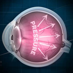
A: Glaucoma is caused by high levels of pressure within the eye. This intraocular pressure (IOP) presses on the optic nerve, causing damage, and eventually leading to permanent vision loss.
Q5: What causes high eye pressure?
A: Pressure builds within the eye as a result of a problem in the eye’s drainage system. In a healthy eye, the aqueous humor, or fluid, is produced within the eye— and as it drains out of the eye through the anterior angle, new fluid is produced. This process repeats itself continuously, and if effective, maintains a normal level of eye pressure.
When a problem occurs within the drainage system, and the eye’s natural drain becomes clogged, the fluid builds up in the eye, and consequently causes a buildup of pressure within the eye.
An easier way to understand how eye pressure increases, is imagining the sink in your kitchen. When the faucet is turned on, the water flows into the basin of the sink and continues down the drain pipe. When there is a blockage in the drain pipe, the water continues to flow into the pipe, causing increased pressure in the pipe.
Just as constant high pressure within the drain pipe will cause damage over time and may lead to a burst pipe, constant high pressure in the eye will eventually lead to optic nerve damage, and consequently vision loss.
Q6: Will glaucoma affect all of my vision?
A: Peripheral vision is affected first, and if not effectively controlled at this point, can result in tunnel vision. As the disease progresses, central vision is affected next, leaving the person with partial, or complete permanent vision loss.
Q7: What is ‘tunnel vision’?
A: If glaucoma is not effectively controlled at an early stage, the disease can cause severe peripheral vision loss— resulting in a condition called ‘tunnel vision’. Tunnel vision blocks your ‘side vision’, and limits your field of vision to strictly seeing images in your central vision, or straight ahead.
Q8: Is there only one type of glaucoma?
A: There are four primary types of glaucoma:
1. Chronic (open angle) glaucoma is the most common form, caused by IOP build-up over time, typically as a result of aging. Open angle glaucoma affects approximately 2.7 million Americans, above the age of 40.
2. Acute (angle closure) glaucoma is a rare form that requires immediate medical attention. It develops as a result of sudden pressure build-up. Symptoms include blurred vision, seeing halos around lights, eye pain, headaches, nausea and vomiting. Acute angle closure glaucoma affects about 1 in 1,000 people.
3. Secondary glaucoma can develop from complications of medical conditions such as, diabetes and hypertension, other ocular conditions, side effects of medications, or trauma to the eye. Secondary glaucoma accounts for 10% of all glaucoma cases.
4. Normal tension glaucoma develops when the optic nerve is damaged, without a presence of high IOP. The cause of this type of glaucoma is still unknown. Normal tension glaucoma accounts for approximately 30-40% of all patients diagnosed with glaucoma.
Q9: How is glaucoma diagnosed?
A: Glaucoma is diagnosed during a comprehensive eye exam, using specific diagnostic tools and assessments:
Tonometry calculates your intraocular pressure (IOP) based on your eye’s response to a puff of air.
Tonometer or tonopen is a tool used to obtain another measure of your IOP. During this test, your eyes will be numbed so that your doctor can gently touch the surface of your eye with either a tonometer or tonopen.
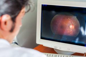
An optical coherence tomography (OCT) scan is a non-invasive, diagnostic tool that uses a laser to provide color-coded, cross sectional images of the retina and optic nerve.
A visual field test produces a computerized map of your visual field and measures the range of your peripheral vision.
Optic disc examination involves a thorough evaluation of the surface of the optic nerves, to examine the quality of the margin, or neuroretinal rim, and optic disc cup.
This examination can be performed with direct ophthalmoscopy, slit lamp, optic disc photography, or with digital imaging devices.
Examination of anterior angle (gonioscopy) enables a thorough evaluation of the anterior angle, the internal drainage system of the eye, located between the iris and cornea.
Through this test, your eye doctor will be able to determine if the angle is open or closed, and detect the presence of abnormal blood vessels, adhesions (synechiae), or damage from previous eye trauma.
To enable a clear view of the angle and its drainage system, your eyes will be numbed, and a special contact lens prism will be placed on the surface of your eye. This test is relatively quick, and does not cause any pain, though you may feel some pressure from the contact lens.
If you suspect you have any eye condition, contact an eye doctor near you, who can diagnose and treat the condition.
SEE RELATED: Normal Tension Glaucoma
Q10: How is glaucoma treated?
A: Unfortunately, a cure for glaucoma and the ability to reverse it’s damage to the eye has yet to be discovered. However, there are treatments available to help stop, or at least reduce the progression of the disease by lowering or controlling IOP:
Anti-glaucoma eye drops are often the first plan of action, as most cases of glaucoma can be controlled with eye drops. Your eye doctor will prescribe a specific type of eye drop, depending on the type of glaucoma.
Types of eye drops:
- Prostaglandins relax the muscles in the eyes to allow better fluid drainage, reducing build-up of IOP.
- Beta-blockers are used in a variety of glaucoma eye drops. They decrease the amount of ocular fluid production and are often prescribed in combination with prostaglandins.
- Alpha-adrenergic agonists decrease the rate of fluid production. They can be used alone or in combination with other anti-glaucoma eye drops.
- Carbonic anhydrase inhibitors decrease the rate of fluid production, and are used in combination with other anti-glaucoma eye drops
- Parasympathomimetics are very often used to control IOP in narrow-angle glaucoma. They work to increase ocular fluid drainage by opening the narrow angle where drainage occurs.
- Epinephrine decreases the rate of ocular fluid production, and increases its outflow from the eye.
- Hyperosmotic agents are used in emergencies, for patients with a severely high IOP that must be reduced immediately before permanent damage occurs. They reduce IOP by lowering ocular fluid volume.
- Combination glaucoma drugs include two different anti-glaucoma medicines. Many times, patients with glaucoma require more than one type of medication to control IOP.
Laser treatment for glaucoma involves creating avenues for increased fluid drainage to reduce IOP.
Selective laser trabeculoplasty (SLT) is the primary laser surgery performed for open angle glaucoma, as it effectively lowers IOP by 20 to 30 percent, and is successful in about 80 percent of patients. This laser procedure stimulates a biochemical change that improves the fluid drainage from the eye. Improved IOP after an SLT treatment may last 3 to 5 years and can be repeated if needed
According to research, the effectiveness of SLT is similar to the most effective glaucoma eye drops.
Other surgery techniques used to treat angle closure glaucoma:
- Iridotomy is performed for angle-closure glaucoma. During this laser procedure, a tiny hole is created in the iris to release the build fluid and allow it to flow properly out of the regular drainage angle.
- Trabeculectomy, is a non-laser procedure that is performed in cases of advanced glaucoma, where there is already optic nerve damage and severe IOP levels. During this procedure, an artificial opening in the eye for ocular fluid drainage is created, to decrease IOP levels.
Q11: What is minimally invasive glaucoma surgery (MIGS)?
A: While many surgeries are often effective in lowering IOP levels, they do pose a risk of potential complications. Minimally invasive glaucoma surgery (MIGS) uses microscopic tools and devices to lower your risk of surgical complications, but to date, can only be performed in combination with cataract surgery.
Micro trabeculectomies involve inserting a microscopic tube into the eye to enable proper fluid drainage through the anterior angle.
There are different devices used for this procedure such as the iStent, Xen Gel Stent and PRESERFLO (formerly called InnFocus and Microshunt).
The iStent® is the most frequently performed MIGS for mild to moderate open angle glaucoma.
The iStent procedure involves implanting a small titanium microtrabecular device to create a bypass through the anterior angle. The iStent enables improved outflow of fluid from the eye, through its natural drainage system— lowering IOP levels.
Benefits of iStent:
- Typically easily combined with cataract surgery— improving IOP with minimal use of pressure lowering eye drops.
- Minimal damage of normal eye tissue.
- Easier to perform, faster to recover from, and safer than many glaucoma surgeries.
- May help reduce a patient’s medication burden— reducing the cost, inconvenience, or side effects of using eye drops.
- May help delay or prevent the need for additional surgical interventions in the future.
Glaucoma filtration surgery
This type of surgery is recommended for early to moderate stages of glaucoma, as the IOP does not reach very low levels through this procedure.
This procedure uses tiny devices and equipment to cut through the trabecular meshwork, without causing harm to any other tissues in the drainage pathway. Using a special contact lens on the eye, a tiny device is inserted into the eye. The trabecular meshwork can either be destroyed with a trabectome or trab360, or bypassed using an iStent. These procedures are FDA-approved but generally don’t reduce the eye pressure to a low enough level for them to be effective in more advanced stages of glaucoma.
Suprachoroidal Shunts
This surgery can successfully lower IOP levels and can be effective in treating moderate to severe glaucoma.
This procedure uses tiny tubes with very small openings, called glaukos shunts, to connect the front of the eye to the suprachoroidal space between the retina and the wall of the eye. The shunt serves to enable proper fluid drainage from the eye.
Q12: Can I prevent glaucoma?
A: While glaucoma cannot be prevented completely, the most reliable way to prevent vision loss from glaucoma is by visiting your eye doctor for regular eye exams. Early detection of the disease can help to preserve your vision, and reduce vision loss.
There are also lifestyle changes you can make to reduce your risk of high eye pressure that leads to glaucoma:
- Eat a healthy diet
- Exercise
- Watch your caffeine intake
- Wear protective eyewear
- Avoid prolonged use of steroids
- Control blood pressure
- Maintain good oral health
LEARN MORE: Guide to Eye Conditions
If you have been diagnosed with glaucoma, or have a high risk of developing this disease, schedule an appointment with an eye doctor for a comprehensive evaluation of your vision and ocular health.