Did you know that the back of the eye is responsible for transferring visual information from the eye to the brain?
In order to see clearly, light is focused onto the back of your eye where it is transformed into electrical impulses. These impulses travel from your eye to your brain, to be processed, organized and interpreted— providing you with an image you can see and understand.
The following ocular structures lie within the deepest layer of the eye, also known as the posterior area of the eye.
Vitreous
The vitreous chamber is located in the back of the eye, and is the largest of the eye’s chambers— accounting for around 80 percent of the eye. (You may remember the anterior chamber, located in the front of the eye).
The vitreous humor, commonly called the vitreous, is a clear jelly-like fluid that fills the space between the lens and the retina.
Most of the vitreous consists of water, while the rest is a combination of collagen, proteins, salts and sugars— all contributing to its gelatinous consistency.
The vitreous is responsible for maintaining the shape of the eyeball, and protecting its inner structures. The vitreous also enables the retina to remain in its place, connected to the back of the eye.
Over time, the vitreous loses its jelly-like consistency and becomes smaller and more liquidy. The collagen and proteins may also form clumps or strings which float around the vitreous, causing shadows to form over the retina. Many people notice these floaters out of the corner of their eyes.
When the vitreous becomes thin, it can cause posterior vitreous detachment, when the retina detaches from the back of the eye. This condition can also cause the appearance of floaters which generally go away over time, and typically do not cause any long term effects.
Sclera
The sclera is the outermost layer of the eye, also known as the white of the eye.
The sclera is made up of tough fibrous tissue and is responsible for giving the eye it’s round shape and protecting the inner structures of the eye.
The sclera consists of three parts:
- Episclera- the loose connective tissue located directly under the conjunctiva
- Sclera proper- the dense white tissue that gives the sclera its white color
- Lamina fusca- the innermost area that consists of elastic fibers
The sclera accounts for more than 80 percent of the eye’s surface area, stretching from the cornea all the way to the optic nerve. This fibrous tissue is thickest in the back of the eye, as it provides extra protection to the area around the optic nerve.
Choroid
The choroid is a thin vascular tissue, located between the sclera and the retina.
This membrane forms the uveal tract, along with the iris and ciliary body, and is responsible for bringing oxygen and nutrients to the outer part of the retina.
The choroid contains four different layers:
- Haller’s layer – contains large blood vessels
- Sattler’s layer- contains medium-size blood vessels
- Choriocapillaris – contains capillaries
- Bruch’s membrane – the innermost layer of the choroid
The choroid also contains a dark-colored pigment, called melanin. Melanin protects the blood vessels in the choroid from toxic light, and absorbs light to prevent light reflections that may interfere with vision clarity— this pigment can sometimes cause the appearance of “red eyes” in flash photography.
This vascular membrane also contains cells that contribute to scleral growth, and its blood flow may help to regulate the temperature within the retina.
If the choroid becomes infected, the macula and optic nerve can be affected as well— many times, causing severe vision loss and even blindness.
When abnormal blood vessels grow from the choroid into the retina, wet macular degeneration can develop.
If you suspect you have an eye condition, contact an eye doctor near you, who can diagnose and treat the condition.
SEE RELATED: Eye Anatomy: The Front of the Eye
Retina
The retina is a thin, light-sensitive membrane that lines the back of the eye.
The retina is responsible for receiving the light that enters the eye, transforming the light into electrical impulses, and sending the visual information to the brain.
The retina has two types of photoreceptors, or light sensitive cells: rods and cones.
The rods are more concentrated on the retina’s periphery, and are essential for discriminating between light and dark colors, detecting movement, and identifying shapes.
The cones are highly concentrated in the center of the retina, within the macula, and are essential for color discrimination and vision for fine details.
Retinal diseases can develop as a result of any type of retinal damage. Untreated retinal diseases can lead to severe vision loss and even blindness. With early detection, some retinal diseases can be treated, while others can be controlled or slowed down to preserve or sometimes restore vision.
Macula
The macula is located in the center of the retina and is responsible for providing sharp, detailed central vision.
This area of the retina contains a high concentration of cone cells, which provide vision for color and detail, and enable sharp central vision.
The fovea is located within the center of the macula and is marked by a small depression. This area consists of highly compacted cone cells that are more narrow and rod-like in shape, when compared to the cones in other areas of the macula.
Due to the high concentration of cone cells, the fovea provides the sharp central vision needed for driving, reading, and other activities that require strong visual acuity.
Macular diseases, including macular degeneration and diabetic eye diseases can occur when macular cells become damaged or destroyed, causing gradual central vision loss.
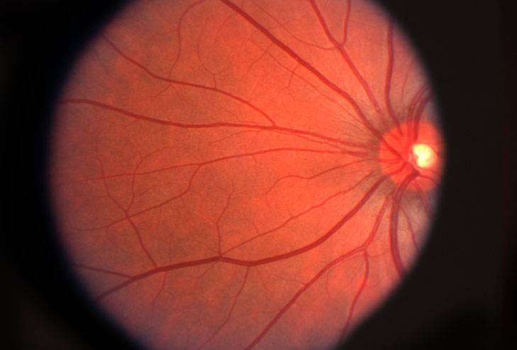
Optic nerve
The optic nerve (cranial nerve II) is a bundle of over one million nerve fibers, located in the back of the eye.
The optic nerve is known as the communication line between the eye and the brain, as it transmits the visual information from the retina to the brain, where it is processed and interpreted.
Since the optic nerve does not contain any photoreceptors, every human has a natural blind spot— though this blind spot is typically not noticed.
The optic disc is the weakest area of the nerve, and is located where the nerve exits the eye.
Glaucoma is one of the most common eye diseases that affects the optic nerve. Glaucoma develops when high eye pressure (intraocular pressure) pushes on the optic nerve, causing permanent nerve damage and resulting in vision loss.
Central retinal artery
The central retinal artery enters the eye through the optic nerve and is responsible for supplying blood to the retina. When this artery enters the eye, it branches out to cover the entire span of the retina.
The central retinal artery is in truth an arteriole, a very thin and fragile blood vessel, and can become easily damaged by an ocular disease, diabetes or any other systemic disease.
Central retinal vein
The central retinal vein is the primary blood vessel that carries the blood out of the eye to travel to the heart and lungs to collect more oxygen. Similar to the central retinal artery, as this vein enters the eye, it branches out to cover the entire span of the retina.
The central retinal vein is also fragile in nature due to its small size, and can be easily damaged by any type of ocular disease.
LEARN MORE: Guide to Eye Health
Schedule an appointment with an eye doctor for a comprehensive eye exam, and to discuss any questions you may have about treating your eye condition.