The external parts of the eye work together to protect the eye and all of its internal structures.
The following ocular structures are located on the eye’s exterior:
Eyelids
The upper and lower eyelids form a moist region around the eye, and protect the surface of the eye from injury, infection, and disease.
The eyelids contain muscles that enable them to open and close around the eye, and are covered with skin. The inner part of the eyelid is lined with mucous membrane, while the outer part is lined with eyelashes.
As the eyelids open and close, they spread the tear film over the eye’s surface, lubricating the eye. The eyelids also remove the tears from the eye’s surface by pushing the tears through the lacrimal puncta into the tear duct, this is called the ‘watermelon seed’ effect.
The edges of the upper and lower lids are lubricated by an oily secretion produced by the meibomian glands, which are located inside the eyelids. This oily secretion contributes to the makeup of the tear film, and serves to decrease the rate at which the tears evaporate.
Eyelid disorders include any type of inflammation, infection, benign and malignant tumors, and structural problems.
Blepharitis is one of the most common ocular conditions that affects the eyelids. It is caused by an inflammation of the eyelids, usually as a result of a blockage in the meibomian glands at the base of the eyelashes. When meibomian glands do not produce enough oil, or produce oil of poor quality, bacterial growth may occur— resulting in a bacterial infection.
If you suffer from any eye conditions related to your eyelids contact an eye doctor near you to help manage your condition.
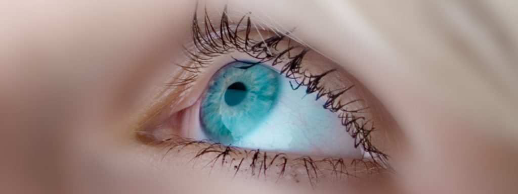
Eyelashes
The eyelashes are the hairs that grow along the edges of the upper and lower eyelids. The eyelashes protect the eye from foreign particles, such as dust, pollen, and debris. The eyelashes are sensitive to touch and send signals to the eyelids to close when a foreign object comes too close to the eye.
The eyelashes fall out on their own and take around six to eight weeks to grow back. Pulling out the eyelashes can cause damage to the eyelash follicles under the skin, from which the hairs grow.
Eyelashes only grow to a certain length, and never need to be trimmed. Eyelash color may differ from hair color, though people with dark hair will typically have darker lashes, while people with light hair will usually have lighter lashes.
Certain ocular conditions, such as blepharitis and demodex mites, that can affect the eyelashes and cause them to become flaky or fall out.
Meibomian glands
Meibomian glands are the oil glands located inside the eyelids. Their opening pores line the edges of the eyelids, near the eyelashes.
Around 25 to 40 meibomian glands line the upper eyelid and 20 to 30 line the lower eyelid. These glands secrete the oil that coats the eye and tear film, to prevent the tears from evaporating too quickly.
Meibomian gland dysfunction (MGD) is a common condition that can develop when the oil glands become blocked with thick secretions, preventing a normal flow of oil and reducing oil quantity and quality. In many cases, this results in dry, irritated eyes and dry eye syndrome.
If you suffer from any eye conditions related to your eyelids contact an eye doctor near you to help manage your condition.
SEE RELATED: Eye Anatomy: The Front of the Eye
Lacrimal gland
The lacrimal gland creates the water layer of the tears that lubricate, cleanse, nourish, and protect the surface of the eye. This gland is located on the upper lateral wall of the eye, under the eyebrow.
When uveitis or an autoimmune disorder affects the lacrimal glands, dry eye syndrome can result.
Tear duct
The tear duct, also called the nasolacrimal duct, is located in the inner corner of the eye, and is part of the tear drainage system that goes from the eye, through the back of the nose, and down the throat.
The tears are spread across the eye’s surface to moisten, clean, nourish, and protect the ocular tissues. When the eyelids open and close, they push the tears out of the eye through the tear duct, to be drained down the back of the nose.
When the tear duct becomes clogged or blocked, the tears are unable to drain out of the eye properly— usually resulting in an irritated, watery eye.
A blocked tear duct is common among newborns, but can also occur in adults as following an eye injury, infection, or as a result of an ocular tumor.
Extraocular muscles
There are six extraocular muscles that attach to the outside of the eye from the bone in the eye’s socket. These muscles work to rotate the eye, and move the eye up, down, and from side to side. When they work together, these muscles can move the eye in any direction.
- Medial Rectus (MR) – Moves the eye inward, towards the nose
- Lateral Rectus (LR) – Moves the eye outward, away from the nose
- Superior Rectus (SR) – Moves the eye upward and inward, and rotates the top of the eye toward the nose
- Inferior Rectus (IR) – Moves the eye downward and inward, and rotates the top of the eye away from the nose
- Superior Oblique (SO) – Moves the eye downward and outward, and rotates the top of the eye toward the nose
- Inferior Oblique (IR) – Moves the eye upward and outward, and rotates the top of the eye away from the nose
Click here to learn more about the ocular structures in the front of the eye.
Click here to learn more about the ocular structures in the back of the eye.
LEARN MORE: Guide to Eye Health
Schedule an eye exam with an eye doctor near you who has experience diagnosing and treating eye conditions.