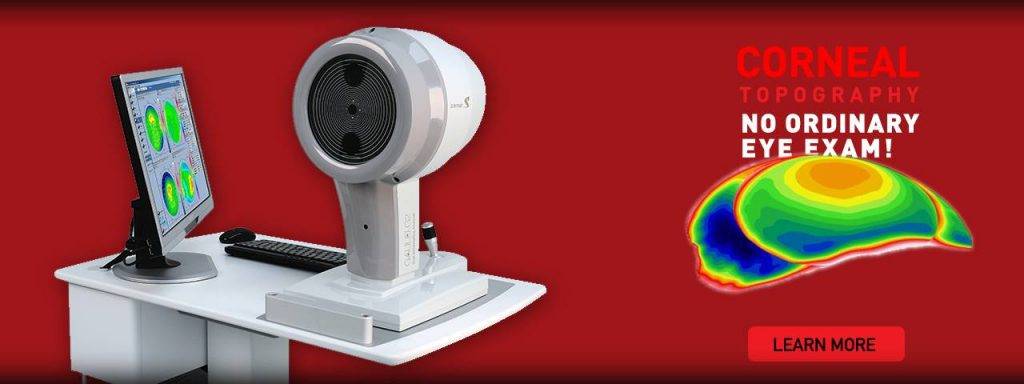
Corneal topography, also known as corneal mapping, is a diagnostic tool that provides 3-D images of the cornea.
The cornea is the outer layer of the eye, responsible for about 70 percent of the eye’s focusing power. A corneal topography test provides detailed 3D maps of the cornea’s shape and curvature and enables detection of corneal diseases, and irregular corneal conditions, such as swelling, scarring, abrasions, deformities, and irregular astigmatisms.
Corneal topography is also used to monitor and treat corneal conditions, plan eye surgery, and fit contact lenses.
Types of corneal topography
There are three different types of technologies used for corneal topography:
Placido disc topography
Placido disc reflection systems measure the curvature, irregularities, tear film quality, foreign bodies, and other parts of the anterior cornea. The reflection is highly dependent on the tear film which reflects the light, and can be either small-cone or large-cone. Small cones are more accurate as they collect more data points, but large cones are easier to use and make collecting data significantly easier.
Scheimpflug and scanning-slit topography
These two systems provide information about the anterior and posterior cornea, and are used for detection and management of corneal swelling, which is specifically important for contact lens wearers.
Types of topographic maps
Axial display map
This is the most traditional way of viewing a topography image, as it is known for its overview of the corneal power. However, since it collects the averages of the data to produce a smooth map, it is considered less accurate than the other maps.
Axial maps are a helpful tool for selecting the base curve of a soft contact lens because the average of the central curvature is displayed. However, for information about corneal shape and power, other map displays are more ideal.
Tangential display map
This type of map provides an accurate measurement of the cornea’s power and curvature, and is therefore helpful in fitting contact lenses, especially ortho-k lenses.
This map can also be used to evaluate the power of a contact lens while the lens is on the eye. This is helpful when the patient is being fitted for a multifocal contact lens and the optical powers must be correctly positioned on the eye.
The tangential map is ideal for detecting changes in the corneal curvature that may occur as a result of corneal distortion from wearing contact lenses.
Elevation display map
This map is used to determine the true shape of the cornea and is crucial for selecting the best contact lens design for an irregular cornea. This map display is especially important when deciding between a scleral gas permeable (GP) lens or a corneal contact lens.
The elevation display map is also helpful for ortho-k management, as corneal shape is crucial in determining if ortho-k lenses will be appropriate for the patient, and in selecting a dual-axis or single-axis lens.
Corneal thickness display map
This map display is used to stage ocular diseases, such as keratoconus, but is primarily used to monitor changes in corneal thickness during contact lens wear.
Tear break-up display
This map displays the quality of the natural tear film and also shows how the tear quality has been impacted by contact lens wear. A corneal topography test will measure the tear film before the patient begins to wear contact lenses and then be compared to the measurement taken after the patient has been wearing contact lenses.
Contact an eye doctor near you who has experience diagnosing and treating corneal conditions.
SEE RELATED: What Is an OCT Eye Exam?
What should I expect during a corneal topography test?
A corneal topography test is quick and painless. During the test, you will sit in front of a lighted bowl that contains a pattern of rings, and rest your head against a bar. A series of data points will be collected, and a color coded image of your corneal shape will be generated on a computer screen.
The images will contain different colors to differentiate elevations— similar to a topographic map of the earth that displays changes in the surface of the land.
When is corneal topography used?
Corneal topography can be used for a variety of reasons:
- Keratoconus
- Planning refractive surgery
- Monitoring ocular health post refractive surgery
- Determining appropriate intraocular lens for cataract surgery
- Evaluating and treating astigmatism post-keratoplasty
- Detecting corneal conditions such as pterygia, corneal scars, and Salzmann nodules
- Monitoring ocular disease
- Assessing integrity of anterior angle for glaucoma patients
- Measuring corneal depth
- Evaluating corneal nerve
- Detecting infectious keratitis
- Examining corneal endothelium
If your doctor recommends a corneal topography test, you can rest assured that this test is non-invasive, quick, and medically necessary.
LEARN MORE: Guide to Eye Exams
Contact an eye doctor near you to discuss any questions or concerns you may have about a corneal topography test, or your ocular health.
Your ocular health is your doctor’s number one concern.